Brain Images of Normal Subjects
■ Research Data and Face Recognition
■ Press coverage of Brain Atlas paper
■ Brain Image Bank Meeting 2014
■ Visiting Students, Summer 2014
News
December 2019 Research Data and Face Recognition
In order to protect the identity of our subjects when we prepare data for inclusion in BRAINS Imagebank we remove not only identifying metadata but also faces and ears from the images.
This recent experiment, described in correspondence to the New England Journal of Medicine(NEJM), illustrates the potential dangers of not defacing the medical images stored in publicly shared research databases.
Identification of Anonymous MRI Research Participants with Face-Recognition Software New England Journal of Medicine
Authors: Christopher G. Schwarz, Ph.D & Others
June 2017 Publication of report on brain image biobanking
This report follows a meeting of international experts from multiple disciplines, all interested in brain image biobanking.
Authors: Susan D. Shenkin, Cyril Pernet, Thomas E. Nichols, Jean-Baptiste Poline, Paul M. Matthews, Aad van der Lugt, Clare Mackay, Linda Lanyon, Bernard Mazoyer, James P. Boardman, Paul M. Thompson, Nick Fox, Daniel S. Marcus, Aziz Sheikh, Simon R. Cox, Devasuda Anblagan, Dominic E. Job, David Alexander Dickie, David Rodriguez, Joanna M. Wardlaw
April 2016 BRAINS Atlases available
BRAINS has developed seven age-specific atlases of T1 brain MRI from 25 to 92 years. They are available for download from BRAINS atlases @ Edinburgh Datashare
January 2016 BRAINS Imagebank paper
A paper about the BRAINS Imagebank was published in NeuroImage.
Authors: Dominic E. Job, David Alexander Dickie, David Rodriguez, Andrew Robson, Cyril Pernet, Mark Bastin, James P Boardman, Alison D. Murray, Trevor Ahearne, Gordon D. Waiter, Roger T. Staff, Ian J. Deary, Susan D. Shenkin, Joanna M. Wardlaw
Summer 2015 Nuffield Student Placement
Eilidh Edmiston was a visiting student on the BRAINS project in summer 2015, as part of a Nuffield Research Placement (http://www.nuffieldfoundation.org/nuffield-research-placements). She piloted and adapted a questionnaire to understand how clinicians use brain image templates, which will inform a wider online survey.
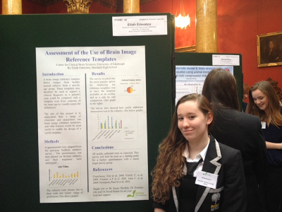
Eilidh presenting her poster at the Royal College of Physicians of Edinburgh.
May 2015 Press coverage of Brain Atlas paper:
David Alexander Dickie , Dominic E. Job, David Rodriguez Gonzalez, Susan D. Shenkin, Joanna M. Wardlaw
Published: May 29, 2015 DOI: 10.1371/journal.pone.0127939
- Interview with BBC Radio Scotland
- The Express
- Eureka Alert
- STV News
- BT Science News
- Consumer Affairs
- The Mirror
- Digital News World
- Science Daily
Brain atlas could help diagnose age-related cognitive impairment
A digital map of the ageing brain could aid the diagnosis of Alzheimer’s disease and other neurodegenerative disorders, a study suggests. The atlas could aid diagnosis by comparing patients’ MRI scans with a map of the healthy ageing brain.Most existing MRI atlases are based on the brains of young and middle-aged people, which don’t reflect the normal changes that take place in the brain as we age, the team says.
Ageing brains
University researchers constructed a detailed atlas of the human brain using MRI scans from more than 130 healthy people aged 60 or over. The team used their atlas to study brain scans taken of normal older subjects and those with Alzheimer’s disease. The atlas was able to pinpoint changes in patients’ brain structure that can be an underlying sign of the condition, researchers say.
Disease signs
A key sign of early Alzheimer’s disease is the loss of brain tissue in a region of the brain, known as the medial temporal lobe. These changes are often subtle and can be difficult to spot, but an MRI atlas could make it easier to detect them, researchers say. The team is continuing to develop MRI atlases of the healthy brain across the lifespan as part of a project - Brain Imaging in Normal Subjects (BRAINS) - which aims to detect brain damage in other diseases such as schizophrenia and preterm birth.
Early Diagnosis
For atlases to be useful and reliable, the team says brain imaging centres need to continue to collect scans from healthy older people and work together to make large brain image banks. The ultimate aim is to use digital brain atlases to support earlier diagnoses of Alzheimer’s and other neurological diseases that develop at different stages of life, the team says. The study, published in the journal PLOS ONE, was principally supported by the Scottish Funding Council, Scottish Imaging Network, A Platform for Scientific Excellence (SINAPSE), and the Medical Research Council.
August 2014 Development of Human Brain Image Banks and Age-Specific Normative Brain Atlases
SFC SINAPSE SPIRIT Programme of Knowledge Exchange in Imaging
The aim of the meeting was to convene a group of world experts in all aspects of creating, curating, and using brain image banks, particularly of normal individuals, to discuss:
- Philosophical and biological issues such as ‘What is normal”, how to facilitate potential clinical and research uses of brain image banks and atlases. Technical issues such as between scanner variation, user access and portals, database infrastructures, statistical issues in image processing and atlas creation, ways to facilitate harmonisation of images and metadata between existing and future planned brain image banks, data sharing, maximization of potential extractable information and impact.
- Legal and ethical issues such as privacy, anonymisation, ‘research tourism’, secondary uses.
- Encourage brain image banks to be compatible world-wide.
- Facilitate ways to contribute imaging collected in publicly funded research or in clinical practice, where appropriate, to brain image banks and made easily accessible.
- Facilitate discussion between image banks in different countries, including to support existing harmonisation initiatives
Suggested articles and resources
Many thanks to our funders whose contributions made this meeting possible:
Summer 2014 Visiting Students
Juyoung Lee and Daniel Taylor were visiting students on the BRAINS project in summer 2014.

Juyoung conducted a systematic review of structural brain MRI atlases. This review documented the subjects and methods used to create structural brain MRI atlases. Initial results show that older subjects (>60 years) were often under-represented and that parametric methods were most commonly used to combine individual images into an atlas.
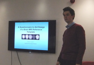
Daniel developed a questionnaire to aid design of a brain MRI atlas interface for supporting radiological diagnoses in individual patients. The questionnaire assessed user preferences and specific features that would be useful in clinical versus research environments. Daniel completed pilot testing of the questionnaire and it will soon be mass distributed electronically to mailing lists of clinicians and researchers.